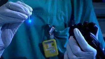
Celiac.com 04/27/2010 - A team of clinicians recently assessed the diagnostic value of confocal endomicroscopy in celiac disease.
Clinicians U. Günther, S. Daum, F. Heller, M. Schumann, C. Loddenkemper, M. Grünbaum, M. Zeitz, and C. Bojarski made up the research team. They are variously affiliated with the Medical Clinic of Gastroenterology, Rheumatology and Infectious Diseases, and with the Department of Pathology, Charité of the Campus Benjamin Franklin of University Medicine Berlin, Germany.
Celiac.com Sponsor (A12):
Studies by Gutschmidt, by Henker and others have shown that, even in the face of greater awareness and more widespread blood testing, a number of countries suffer from low rates of celiac disease detection.
Patchiness of the mucosal and submucosal changes associated with celiac disease impedes proper celiac diagnosis, and contributes to a small, but important sampling error among those assessed.
Some clinicians feel that the problem might be resolved somewhat by modifying the endoscopic screening process to include real-time assessment of the duodenal morphology. Better endoscopic and imaging procedures will mean better diagnostic accuracy of biopsy specimens.
One early step toward improved gathering of targeted biopsies in celiac disease patients lies in using magnification endoscopy, either alone, or together with high-resolution narrow-band imaging. This method offers better ability to detect patchy villous atrophy sites in the duodenum.
However, neither magnification, nor narrow-band imaging techniques can detect increased number of intraepithelial lymphocytes (IELs).
Confocal endomicroscopy (CEM) permits live, real-time histology with a 1000x magnification during endoscopy. Recently, a prospective pilot study for the first time showed CEM to enable effective live, real-time diagnosis and evaluation of celiac disease. Researchers compared endomicroscopic findings during live, real-time endoscopy with histological findings graded according to Marsh classification.
The research team used CEM to examine twenty-four patients with celiac disease and six patients with refractory celiac disease, all following a gluten-free diet.
The team evaluated duodenal mucosa by CEM and by conventional histological analysis for villous atrophy, crypt hyperplasia, and increased numbers of intraepithelial lymphocytes (IELs > 40/100 enterocytes).
They then compared the CEM to the histology results for sensitivity, specificity, and inter-observer variability using Marsh classification scores.
As control subjects, the team used thirty patients without celiac disease, but who were undergoing routine upper gastrointestinal endoscopy.
Using conventional histology on the 30 patients with celiac disease, the team found 23 cases with villous atrophy and crypt hyperplasia, and 27 cases of increased IELs.
Using CEM, the team found 17 cases of villous atrophy, 12 cases of crypt hyperplasia, and 22 cases of increased IELs. The CEM results compared favorably to those of conventional histology for villous atrophy, for which CEM showed a sensitivity of 74%, and for increased numbers of IELs, for which CEM showed a sensitivity of 81%. However, the CEM results were lacking for detecting crypt hyperplasia, for which CEM showed a sensitivity of 52%.
The κ values for determination of inter-observer variability were 0.90 for villous atrophy, 1.00 for crypt hyperplasia, and 0.84 for IEL detection. For the 30 control patients, both traditional histology and CEM revealed normal duodenal architecture with overall specificity of 100%.
From these results, the team concluded that the assessment of duodenal histology by CEM in patients with celiac disease is sensitive and specific in determining increased numbers of IELs and villous atrophy, but inadequate for detecting crypt hyperplasia. Until improvements are made, CEM is not suitable for use in diagnosing celiac disease.
SOURCE: Open Original Shared Link


Recommended Comments
There are no comments to display.