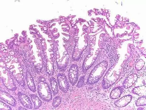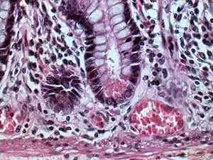
Celiac.com 05/05/2010 - Celiac disease is the most commonly misdiagnosed auto-immune disease of modern times. People that have celiac disease and ingest gluten, have a T-Cell mediated immune reaction which creates damage to the small intestinal mucosa. Mucosa villous atrophy is presented as an abnormality of the small intestine, and results in the flattening of the mucosa, and gives the appearance of atrophy of villi. Clinically, this is found in malabsorbtion syndromes like celiac disease. The degree of damage which occurs in the duodenum can vary, and there is some controversy regarding the coexistence of villous atrophy and normal mucosa found in different biopsy locations.
Tests for villous atrophy were conducted at the regional referral center Gastrointestinal Patho-physiology and Endoscopy, University Department of Pediatrics, Children's Hospital, Spedali Civili, Brescia, Italy; and University Department of Pathology II, Spedali Civili, Brescia, Italy, on all children below 2 years of age and all patients with positive serum IgA antibodies or raised serum IgG anti-gliadin antibodies. The central focus of the study was to analyze the variability anddistribution patterns of histological lesions in celiac children. Eachbiopsy taken was thoroughly analyzed, and each type of lesion found wasdocumented.
Celiac.com Sponsor (A12):
Six hundred and eighty-six children enrolled as patients at the clinic between July 2005 and October 2009, tested positive for celiac disease. Of the 686 celiac patients, none of them had an entirely normal biopsy, 96.2% had some degree of villous atrophy, 80.1% had total villous atrophy, and 46.6% had different lesions in different places. 16.9% or 116 of the patients studied had lesions that varied within the same biopsy. Of those 116 patients, all of them also had histological normal lesions within the same biopsy.
There was no determined correlation between distribution and type of histological lesions and medical presentation of celiac disease. In the 800 children with celiac that were evaluated for this and previous studies, there was absolutely no evidence of of any cases where any lesions, including villous atrophy were isolated to the duodenal bulb. Additionally, there were no biopsy's where the intraepithelial lymphocyte (IEL) count was normal, indicating that there is no truly normal duodenal histology in celiac patients.
Some variability of histological lesions were found even within the same duodenal biopsy. Not only did this study confirm that duodenal lesions can vary among varying biopsies, it also demonstrated that severity of lesions has a proximal-to-distal gradient, but no patient has a completely normal duodenal biopsy. This discovery of histological variation in celiac biopsy's may help to establish an accurate celiac diagnosis for celiac in the future.
Source:
- Open Original Shared Link


Recommended Comments
There are no comments to display.