Celiac.com 04/16/2021 - When undergoing an evaluation for possible Celiac disease or gluten sensitive enteropathy doctors usually recommend an upper endoscopy with small intestinal biopsy. What that is and why it is recommended may not be clear to many people who are facing the decision of whether to undergo the procedure themselves or whether to subject their child to this exam.
What is an endoscopy and how is it done?
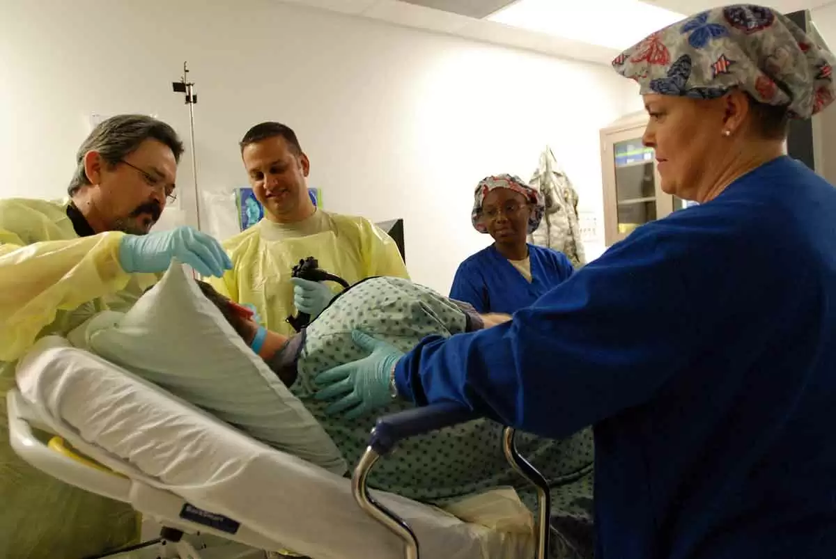
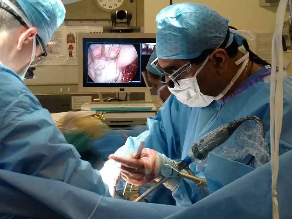
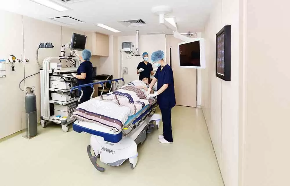
During upper endoscopy a thin flexible tube, about the diameter of a fat pencil, is passed in the mouth down the upper gastrointestinal tract. This tube has a video chip on its tip. From the mouth it is advanced down the esophagus, or inside a feeding tube, into the stomach. It is then advanced into the first part of the small intestine known as the duodenum. Thus, upper endoscopy is also known as esophagogastroduodenoscopy or EGD for short. The scope has internal channels for flushing water, suctioning secretions and passing instruments that can obtain pinch biopsy samples of tissue for microscopic examination. The scope dials control internal cables that allow the tube to be turned up/down and right/left at the tip.
Do you feel and endoscopy or remember it?
Celiac.com Sponsor (A12):
Typically, people undergoing the exam in the U.S. are sedated with medication. These medications are similar to Valium. They have a good amnesia and relaxing effect and are called midazolam or versed and are usually combined with a narcotic pain medication like meperidine (Demerol) or Fentanyl. The result is a sort of drowsy twilight amnesia. Lately, a very short-acting intravenous sedative, called propofol (diprovan) is increasingly being used for deeper sedation or general anesthesia. Occasionally, usually in very young children or people with severe lung problems, general anesthesia is required. The exam is usually not felt or remembered because of the medications.
What is examined and how well is the lining is seen?
Celiac disease affects the upper portion of the small intestine, in the two sections known as the duodenum and jejunum. The examination of the small intestine is usually limited to the first section, termed the duodenum, though occasionally the second section known as the jejunum may be reached especially when a longer endoscope is used. The video images are very high resolution with the latest endoscopes and may have a magnification and color contrast mode to detect very subtle signs of damage of the small intestine.
What are the typical endoscopy findings?
The characteristic appearance of the surface of the small intestine in celiac disease includes superficial ulcerations that are commonly linear, flattening of the folds, notching or scalloping of the folds and a mosaic-like pattern. However, the surface may appear normal and only under microscopic examination of samples will the lining show signs of gluten-induced injury.
What are intestinal biopsies? What are their limitations? What can be missed?
Samples of small intestine are obtained with biopsy forceps that consist of tiny jaws with cups that permit pinching off samples of the intestinal lining. This is painless and very safe. The samples are placed in a preservative solution and sent to a pathology lab. The tissue is then processed, embedded in paraffin wax, cut into thin slices and mounted on a microscope slide. The slides are stained before being examined under the microscope by a pathologist. Small intestine injury from gluten may be patchy. Therefore, several samples are recommended. A minimum of 4 pieces and preferably 8 to 12 samples should be obtained to avoid missing microscopic signs of Celiac disease.
What does the pathologist look for on the slides to determine if there is Celiac disease or gluten injury?
The pathologist examines the slide for evidence of damage or injury characteristic of gluten sensitivity. Occasionally special stains are required to see signs of irritation known as inflammation. Inflammation in the gut is characterized by an increased number of a type of white blood cells. For Celiac disease the characteristic white blood cell involved in gluten-induced intestinal injury are called lymphocytes. In early celiac and gluten sensitivity without celiac disease, the biopsy may be normal and the diagnosis may not be established by intestinal biopsy without special stains or, in the research setting, by electron microscopy.
Summary
The procedure of endoscopy is safe, painless, and very helpful for confirming the diagnosis of celiac disease while excluding other upper intestinal disorders. However, the main drawback of endoscopy is that nearly everyone must have sedation to tolerate the exam and it can be expensive if not fully covered by the patient’s insurance. Furthermore the biopsies may not confirm the diagnosis. If the biopsy is normal, misread as normal, or is borderline, the diagnosis of celiac disease is neither confirmed nor excluded.
Sometimes, celiac disease is diagnosed by endoscopic biopsy in people who either have normal blood tests or as an incidental finding in those undergoing endoscopy for other reasons. When the biopsy is abnormally classic for Celiac disease and the blood tests are negative but the patient responds to a gluten-free diet, the term seronegative (blood test negative) celiac disease is often used. Fear or confusion about endoscopy should not prevent anyone who is suspected of having celiac or gluten sensitivity from undergoing endoscopy.


Recommended Comments
There are no comments to display.
Create an account or sign in to comment
You need to be a member in order to leave a comment
Create an account
Sign up for a new account in our community. It's easy!
Register a new accountSign in
Already have an account? Sign in here.
Sign In Now