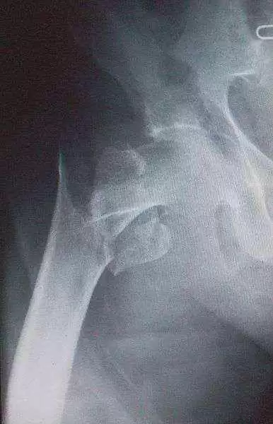
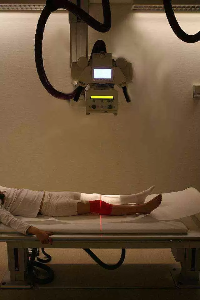
Celiac.com 10/26/2015 - Patients with active celiac disease are more likely to have osteoporosis and a higher risk of bone fractures. High-resolution peripheral quantitative computed tomography (HR-pQCT) permits three-dimensional exploration of bone micro-architectural characteristics measuring separately cortical and trabecular compartments, and gives a more profound insight into bone disease pathophysiology and fracture.
 A research team recently assessed the volumetric and micro-architectural aspects of peripheral bones-distal radius and tibia-in an adult premenopausal cohort with active celiac disease assessed at diagnosis. The research team included MB Zanchetta, F Costa, V Longobardi, G Longarini, RM Mazure, ML Moreno, H Vázquez, F Silveira, S Niveloni, E Smecuol, MdeL Temprano, HJ Hwang, A González, EC Mauriño, C Bogado, JR Zanchetta, and JC Bai. They are variously affiliated with IDIM, Instituto de Diagnóstico e Investigaciones Metabólicas, Buenos Aires, Argentina, the Sección Intestino Delgado, Departamento de Medicina, Hospital de Gastroenterología "Dr. C. Bonorino Udaondo", Buenos Aires, Argentina; and the Cátedra de Gastroenterología Facultad de Medicina, Universidad del Salvador, Buenos Aires, Argentina.
Celiac.com Sponsor (A12):
For their study, the team prospectively enrolled 31 consecutive premenopausal women, between 18-49 years of age, with newly diagnosed celiac disease, and 22 healthy women of similar age and body mass index.
Compared with controls the peripheral bones of celiac disease patients showed significantly lower total density mg/cm(3). Celiac patients also showed significantly lower cortical densit in both regions.
Although celiac patients also showed lower cortical thickness, there was no significant inter-group difference (a-8% decay with p 0.11 in both bones). The 22 patients with symptomatic celiac disease showed a greater bone micro-architectural deficit than those with subclinical, or "silent" celiac disease.
The team used HR-pQCT identify significant deterioration in the micro-architecture of trabecular and cortical compartments of peripheral bones. Overall, impairment was marked by lower trabecular number and thickness, which increased trabecular network heterogeneity, and lower cortical density and thickness.
The team notes that they expect a follow-up on this group of patients to reveal whether a gluten-free diet promotes bone healing, and if so, to what extent.
Source:
- Open Original Shared Link


Recommended Comments