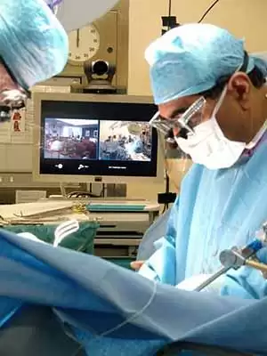
Celiac.com 01/11/2006 - For many years, biopsy of the small bowel demonstrating villous atrophy has been fundamental to the diagnosis of celiac disease. Older celiacs will remember, fondly or otherwise, the Crosby suction biopsy device which was swallowed attached to a long tube and made its way down to the small bowel where, position confirmed by x-rays, it guillotined a small portion of tissue. The procedure was tedious and technical failures common—only identified when the device was hauled up after several hours. Later it became clear that biopsies from the duodenum obtained during endoscopy were just as good, and the biopsy process became a five minute job with no need for X-rays. Nevertheless, many celiacs are reluctant to undergo biopsy and its necessity is increasingly questioned, particularly now that blood tests for celiac-related antibodies are highly sensitive and specific. There are a number of reasons why, in my own practice, biopsies continue to be helpful in celiacs diagnosed in adulthood.
- Biopsies are necessary when blood tests are negative. While endomysial (EmA) and tissue transglutaminase (TTGA) antibodies are detectable in most cases where villous atrophy is present, 5-10% of patients lack these antibodies1. In this situation, where the story is suggestive of celiac, perhaps with a family history or strongly suggestive symptoms, biopsy is the only way to make the diagnosis. Increasingly, physicians recognize that many patients with gluten sensitivity do not have villous atrophy (Grade III of the Marsh classification) of "classic" celiac disease, but have milder abnormalities such as crypt hyperplasia (Marsh II) or an excess of the inflammatory cells called lymphocytes (Marsh I). Patients in these categories are less likely to have positive serology2.
- Biopsies are necessary where false positive blood tests may occur. TTGA, particularly where levels are low, may be associated with diseases other than celiac: ulcerative colitis, Crohns disease, arthritis and liver diseases without any evidence of celiac disease have been linked3. Newer TTGA tests have steadily improved in this regard but I still would be reluctant to diagnose celiac on a TTGA test alone. "False positive" EmA is a different issue which I will return to.
- Biopsies give a baseline for comparison. Suppose a patient starts a gluten-free diet without biopsy—we dont know whether she or he had Marsh I, II or III or even normal histology. A year later, same patient develops new symptoms of diarrhea, weight loss, whatever. Well get a duodenal biopsy as part of the workup, but its going to be difficult to interpret without knowing what things were like before going gluten-free. Specifically, a baseline to look back at tells us whether the small bowel is better, worse or no different, and helps us decide whether we need to focus on celiac disease as the most likely cause of new problems or explore other possibilities involving the rest of the gut. The biggest diagnostic disaster of all, of course, is the gluten-free diet started without any sort of baseline investigation including antibodies, raising the specter of the infamous gluten challenge if a definitive diagnosis is needed.
- Biopsies provide a "gold standard" assessment of the state of the bowel. There has been much excitement recently about capsule endoscopy, a wireless device the size of a large pill (not to be confused with the Crosby capsule!) which makes its own way down the small bowel taking pictures as it goes. Characteristic abnormalities can be seen in celiac disease, raising the possibility that this device might be useful in diagnosis. If experience with conventional endoscopes is any guide, however, these abnormalities are missing in a sizeable minority of celiacs particularly with mild disease4 (Capsule endoscopy in its present state of development can not take biopsies). Certainly the capsule allows assessment of the bowel beyond the reach of conventional "anaconda-style" endoscopes, but I am not convinced at present that it can replace biopsy.
- A follow-up biopsy gives an indicator of progress. I offer my patients a repeat biopsy after two years gluten-free and perhaps surprisingly most take up the offer and are keen to hear how things have improved. Ive increased the biopsy interval from one to two years because only 40% of people had complete recovery after 12 months gluten-free5. EmA and TTGA disappearance is only a marker of how successful gluten exclusion has been and is not a reliable indicator of bowel recovery. Does persisting villous atrophy matter if the patient is doing well on a gluten-free diet? Intuitively, one might like to keep a closer eye on the patient with persistently flat biopsies, who could be at greater risk of complications in the future6.
- The endoscopy not only allows examination and biopsy of the duodenum but also a look at the esophagus and stomach. Sad fact of the ageing process is that you start to collect diseases like trading cards, and just because youre celiac doesnt mean you cant have something else. Its important to have a good look for bleeding lesions in the upper gut even if the blood work for a seventy year old with anemia says celiac (and check out the colon too, but thats a topic for another day).
On the other hand, we recognize that biopsies are not always the final arbiter in diagnosis. While the jury is still out on what a TTGA positive, biopsy negative result means with regard to gluten sensitivity, there is plenty of evidence that a positive EmA generally does mean that biopsy abnormalities will follow: My own follow-up of EmA positive, biopsy negative patients indicates that they will develop abnormal histology if not treated7. So it makes sense to start EmA positive people on gluten-free without waiting for significant bowel damage—and as already stated, even a normal baseline biopsy will provide a reference for any problems that might arise in the future.
Celiac.com Sponsor (A12):
Sometimes I meet a patient with bad gut symptoms but completely normal blood work up and biopsies and when all else fails I will run a trial of gluten-free. It often works, particularly if there is a family history of celiac. But then again, if it doesnt, we have a baseline normal biopsy to say there is no need to persevere.
I guess in the future diagnosis of gluten sensitivity will rely on totting up various factors, none individually essential: blood tests, biopsies, family history, genetic testing for the HLA celiac genes. Some researchers are making a case for dropping the biopsy requirement if the antibody blood work checks out in children8, for whom (and for the parents) endoscopy and biopsy is a major issue. In adults however it is quick, straightforward and safe and will remain a key part of my celiac workup.
William Dickey is a gastroenterologist at Altnagelvin Hospital, Londonderry, Northern Ireland, with over 400 celiac patients attending his clinics. His interest in celiac disease goes back some fourteen years and he has published extensively on the subject. He is an associate member of Coeliac UKs Medical Advisory Council.
References:
- Dickey W, McMillan SA, Hughes DF. Sensitivity of serum tissue transglutaminase antibodies for endomysial antibody positive and negative coeliac disease. Scand J Gastroenterol 2001; 36: 511-4.
- Wahab PJ, Crusius JBA, Meijer JWR, Mulder CJJ. Gluten challenge in borderline gluten-sensitive enteropathy. Am J Gastroenterol 2001; 96: 1464-69.
- Di Tola M, Sabbatella L, Anania MC, Viscido A, Caprilli R, Pica R, Paoluzi P, Picarelli A. Anti-tissue transglutaminase antibodies in inflammatory bowel disease: new evidence. Clin Chem Lab Med. 2004;42(10):1092-7.
- Oxentenko AS, Grisolano SW, Murray JA, Burgart LJ, Dierkhising RA, Alexander JA. The insensitivity of endoscopic markers in celiac disease. Am J Gastroenterol. 2002 Apr;97(4):933-8.
- Dickey W, Hughes DF, McMillan SA. Disappearance of endomysial antibodies in treated celiac disease does not indicate histological recovery. Am J Gastroenterol 2000; 95: 712-4.
- Meijer JWR, Wahab PJ, Mulder CJJ. Histologic follow-up of people with celiac disease on a gluten-free diet: slow and incomplete recovery. Am J Clin Pathol 118(3):459-63, 2002 Sep.
- Dickey W, Hughes DF, McMillan SA. Patients with serum IgA endomysial antibodies and intact duodenal villi: clinical characteristics and management options. Scand J Gastroenterol 2005: in press
- Barker CC, Mitton C, Jevon G, Mock T.Can tissue transglutaminase antibody titers replace small-bowel biopsy to diagnose celiac disease in select pediatric populations? Pediatrics. 2005 May;115(5):1341-6


Recommended Comments
Create an account or sign in to comment
You need to be a member in order to leave a comment
Create an account
Sign up for a new account in our community. It's easy!
Register a new accountSign in
Already have an account? Sign in here.
Sign In Now